Home
Uncategories
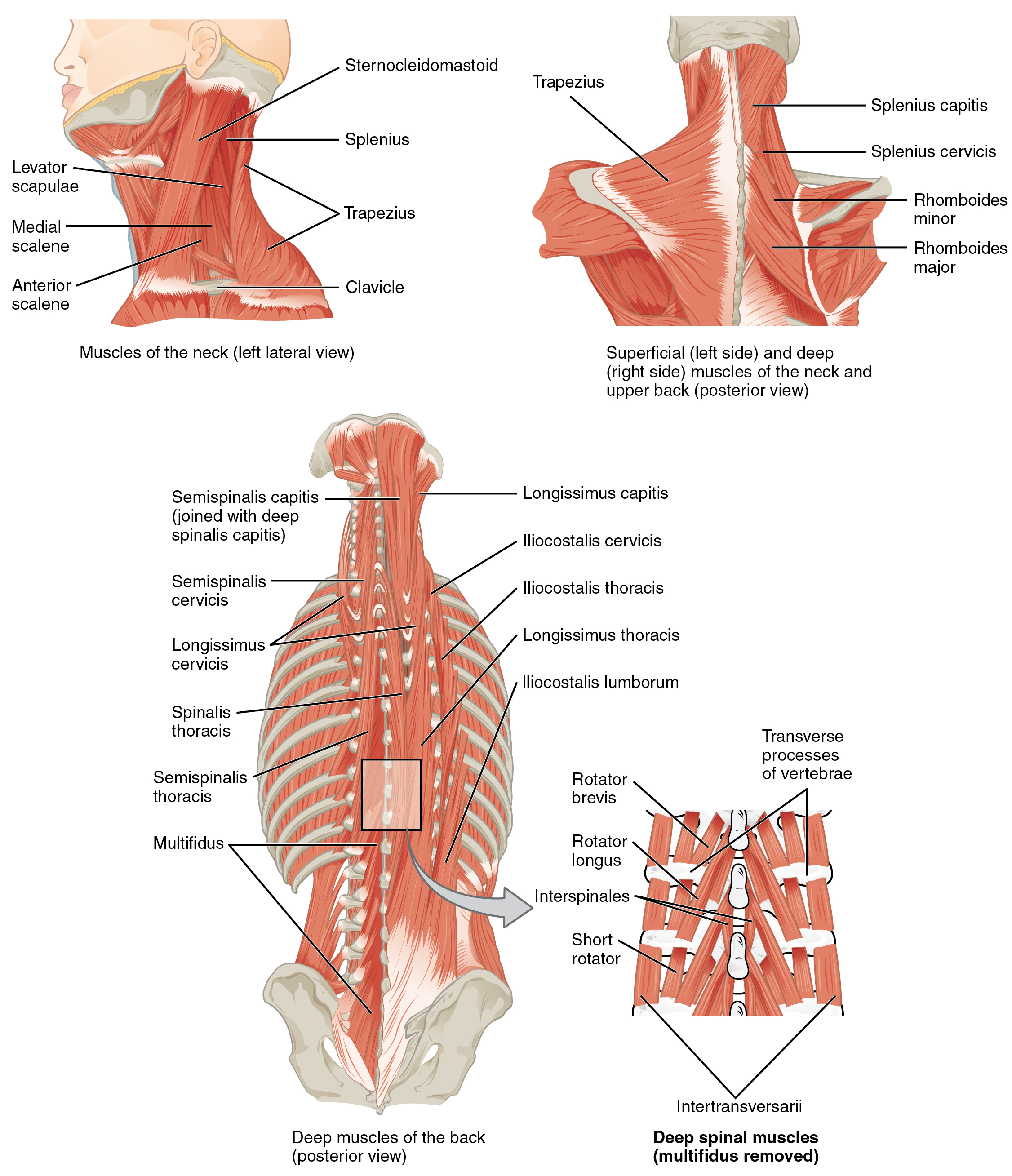
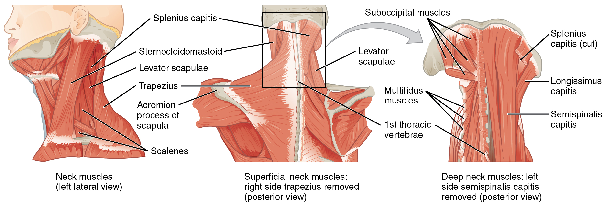
Human Neck Muscle Diagram - The Ventral Neck Muscles Lecturio Online Medical Library - The longissimus (red, in the image above) are located between spinalis and the iliocostalis muscles.
Human Neck Muscle Diagram - The Ventral Neck Muscles Lecturio Online Medical Library - The longissimus (red, in the image above) are located between spinalis and the iliocostalis muscles.
Human Neck Muscle Diagram - The Ventral Neck Muscles Lecturio Online Medical Library - The longissimus (red, in the image above) are located between spinalis and the iliocostalis muscles.. The content of the neck is grouped into 4 neck spaces, called the compartments. Contain the common carotid artery, internal. The muscle anatomy of the head and neck is a fascinating area, with the the neck also containing the 7 vertebrae of the part of the spine called the cervical curve. Photos head muscle diagram anatomy human diagram. The human back extends from the buttocks to the posterior portion of the neck and shoulders.it is opposite from the chest, and the vertebral column runs down the back.
Human muscle system, the muscles of the human body that work the skeletal system, that are under voluntary control, and that are concerned with movement, posture, and balance. Audiologist, speech therapist & cochlear implant specialist. The muscular system makes up nearly half the weight of the human body, this is why when we train we sometimes put on weight instead of losing it. 3d human skeletal system diagram. Attached to the bones of the skeletal system are about 700 named muscles that make up roughly half of a person's body weight.
Axial Muscles Of The Head Neck And Back Anatomy And Physiology from opentextbc.ca Neck diagram human… continue reading → The muscular system is responsible for the movement of the human body. A body muscle diagram is used by different people for various uses. Many in the neck help to stabilize or move the head. There are many muscles around the neck that help to support the cervical spine and allow you to move your head in different directions. The muscular system makes up nearly half the weight of the human body, this is why when we train we sometimes put on weight instead of losing it. The human back extends from the buttocks to the posterior portion of the neck and shoulders.it is opposite from the chest, and the vertebral column runs down the back. Muscles stretch across joints to link one.
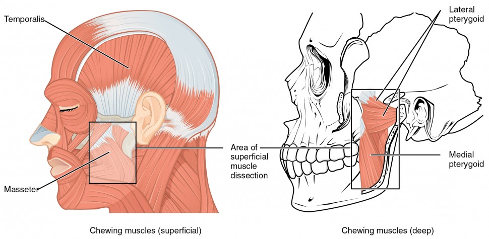
The neck muscles, including the sternocleidomastoid and the trapezius, are responsible for the gross motor movement in the muscular system of the head and neck.
These diverse tasks require both strong, forceful movements and some of the fastest, finest, and most delicate adjustments in the entire human body. There are three sets of longissimus muscles: While classified as peripheral nerves, the motor cell body resides in the anterior horn of the spinal cord. Neck diagram human human lymphatics neck diagram anatomy of diagram. Neck neck muscle anatomy muscle diagram inspirational medical. The muscles of the neck run from the base of the skull to the upper back and work together to bend the head and. Anatomy face and neck side view 12 photos of the anatomy face and neck side view face neck glands, face neck muscles, face neck muscles diagram, face neck muscles picture, face neck pain, human anatomy, neck, face neck glands, face neck muscles, face neck muscles diagram, face neck muscles picture, face neck pain And the lumbrical muscles arising from the. Human neck muscles diagram neck muscle anatomy health medicine and anatomy reference. The back consists of the spine, spinal cord, muscles, ligaments, and nerves. The quizzes below each include 15 multiple choice identification questions related to the muscles of the head and neck. Human leg muscles diagram leg muscle chart gosutalentrankco. 1) above the cervical area (longissimus capitis), 2) in the cervical area (longissimus cervicis), and 3) in the upper back or thoracic area (longissimus thoracis).
Muscles stretch across joints to link one. Muscle head anatomy vocal organ diagram female neck anatomy neck wireframe head neck human anatomy head artery anatomy face pharynx vector neck degree head anatomy 3d. The pelvis at the bottom of the back and the shoulders at the top of the back give the back its breadth, and it narrows in between these two regions. Each of these muscles is a discrete organ constructed of skeletal muscle tissue, blood vessels, tendons, and nerves. These structures work together to support the body, enable a range of movements, and send messages from the brain to.
The Ventral Neck Muscles Lecturio Online Medical Library from d3uigcfkiiww0g.cloudfront.net The muscular system consists of all the muscles present in a single body. Many in the neck help to stabilize or move the head. Neck diagram human human lymphatics neck diagram anatomy of diagram. Here is a list of the many muscles that exist in the neck. The back consists of the spine, spinal cord, muscles, ligaments, and nerves. The content of the neck is grouped into 4 neck spaces, called the compartments. The human muscle diagram provided above is the finger muscle diagram. 3d human skeletal system diagram 13 photos of the 3d human skeletal system diagram human digestive system diagram, human muscular system diagram, human nervous system diagram, skeletal system blank diagram, skeletal system diagram for kids, skeletal system diagram unlabeled, human anatomy, human digestive.
Muscles stretch across joints to link one.
3d human skeletal system diagram. Human neck muscles diagram neck muscle anatomy health medicine and anatomy reference. The muscles of the human body can be categorized into a number of groups which include muscles relating to the head and neck, muscles of the torso or trunk, muscles of the upper limbs, and muscles of the lower limbs. Contain the common carotid artery, internal. The muscle anatomy of the head and neck is a fascinating area, with the the neck also containing the 7 vertebrae of the part of the spine called the cervical curve. Cervical nerves are spinal nerves that arise from the cervical region of the spinal cord. Neck diagram human diagram of the cutaneous nerves of the head and neck stock photo. These diverse tasks require both strong, forceful movements and some of the fastest, finest, and most delicate adjustments in the entire human body. Anterior, lateral and posterior groups, based on their position in the neck.the musculature of the neck is further divided into more specific groups based. Each of these muscles is a discrete organ constructed of skeletal muscle tissue, blood vessels, tendons, and nerves. Neck muscles are bodies of tissue that produce motion in the neck when stimulated. Human muscle system, the muscles of the human body that work the skeletal system, that are under voluntary control, and that are concerned with movement, posture, and balance. The quizzes below each include 15 multiple choice identification questions related to the muscles of the head and neck.
Contain the common carotid artery, internal. Contains glands ( thyroid, parathyroid, and thymus ), the larynx, pharynx and trachea. Neck diagram human… continue reading → These structures work together to support the body, enable a range of movements, and send messages from the brain to. The pelvis at the bottom of the back and the shoulders at the top of the back give the back its breadth, and it narrows in between these two regions.
Axial Muscles Of The Head Neck And Back Anatomy And Physiology I from s3-us-west-2.amazonaws.com 1) above the cervical area (longissimus capitis), 2) in the cervical area (longissimus cervicis), and 3) in the upper back or thoracic area (longissimus thoracis). Contains glands ( thyroid, parathyroid, and thymus ), the larynx, pharynx and trachea. In other positions, other actions may be. Audiologist, speech therapist & cochlear implant specialist. These structures work together to support the body, enable a range of movements, and send messages from the brain to. Skeletal muscles, smooth muscles, cardiac muscles. Many in the neck help to stabilize or move the head. We all have a layer of fatty tissue under our skin, and this softens the look of the underlying.
See anatomy of the head and neck stock video clips.
Cervical nerves are spinal nerves that arise from the cervical region of the spinal cord. See anatomy of the head and neck stock video clips. There are eight pairs of cervical nerves, denoted c1 to c8. The muscles of the human body can be categorized into a number of groups which include muscles relating to the head and neck, muscles of the torso or trunk, muscles of the upper limbs, and muscles of the lower limbs. Anatomy face and neck side view 12 photos of the anatomy face and neck side view face neck glands, face neck muscles, face neck muscles diagram, face neck muscles picture, face neck pain, human anatomy, neck, face neck glands, face neck muscles, face neck muscles diagram, face neck muscles picture, face neck pain There are three sets of longissimus muscles: The interossei muscles (four dorsally and three volarly) originating between the metacarpal bones; The muscle anatomy of the head and neck is a fascinating area, with the the neck also containing the 7 vertebrae of the part of the spine called the cervical curve. Human leg muscles diagram leg muscle chart gosutalentrankco. The longissimus (red, in the image above) are located between spinalis and the iliocostalis muscles. The longissimus (red, in the image above) are located between spinalis and the iliocostalis muscles. Human muscle system, the muscles of the human body that work the skeletal system, that are under voluntary control, and that are concerned with movement, posture, and balance. The human body has three different types of muscles.
We all have a layer of fatty tissue under our skin, and this softens the look of the underlying neck muscle diagram. The content of the neck is grouped into 4 neck spaces, called the compartments.
0 Comments:
Post a Comment